By John Trader, Director of Communications, M2SYS Technology
One of the biggest recurring issues that the biometrics industry faces is that the technology is often misunderstood, in turn perpetuating resistance to its use and creating assumptions based on a general lack of knowledge.
Partially the fault of the industry itself for not being thorough enough on educating consumers about the differences in biometric modalities, there is no better evidence of the continued confusion around the technology than the idea that iris and retina biometrics are one in the same. Let me take a moment to explain the distinct differences between these two, unique forms of biometric identification.
In biometrics, iris and retinal scanning are known as “ocular-based” identification technologies, meaning they rely on unique physiological characteristics of the eye to identify an individual. Even though they both share part of the eye for identification purposes, these biometric modalities are quite different in how they work.
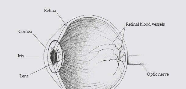
The human retina is a thin tissue composed of neural cells located in the posterior portion of the eye. Because of the complex structure of the capillaries that supply the retina with blood, each person’s retina is unique. The network of blood vessels in the retina is so complex that even identical twins do not share a pattern. Although retinal patterns may be altered in cases of diabetes, glaucoma or retinal degenerative disorders, the retina typically remains unchanged from birth until death.
A biometric identifier known as a retinal scan is used to map the unique patterns of a person’s retina. The blood vessels within the retina absorb light more readily than the surrounding tissue and are easily identified with appropriate lighting.
A retinal scan is performed by casting an unperceived beam of low-energy infrared light into a person’s eye as they look through the scanner’s eyepiece. This beam of light traces a standardized path on the retina. As retinal blood vessels are more absorbent of this light than the rest of the eye, the amount of reflection varies during the scan. The resulting pattern of variations is converted to computer code and stored in a database.
The iris is a thin, circular structure in the eye that is responsible for controlling the diameter and size of the pupil and thus the amount of light reaching the retina. Iris recognition is an automated method of biometric identification that uses mathematical pattern recognition techniques on video images of the irises of an individual’s eyes. These subsequent random patterns are unique and can be seen from some distance.
Unlike retinal scanning, iris recognition uses camera technology with subtle infrared illumination to acquire images of the intricate structures of the iris. Digital templates encoded from these patterns by mathematical and statistical algorithms enable positive identification of an individual. Databases of enrolled templates are searched by matching engines at speeds measured in millions of templates per second and with infinitesimally small false match rates.
Hundreds of millions of people around the world have been enrolled in iris recognition systems for convenience and security purposes from passport-free automated border crossings to national ID functions. A key advantage of iris recognition, besides its speed of matching and its extreme resistance to false matches, is the stability of the iris as an internal, protected, yet externally visible organ of the eye.
While both iris and retina scanning are ocular-based biometric technologies, there are distinct differences that clearly separate the two modalities. Iris Recognition uses a camera, similar to any digital camera, to capture an image of the Iris. The Iris is the colored ring around the pupil of the eye and is the only internal organ visible from outside the body. This allows for a non-intrusive method of capturing an image since you can simply take a picture of the iris from some distance.
Retinal scanning, on the other hand, requires a very close encounter with a scanning device that sends a beam of light deep inside the eye to capture an image of the retina. Since the retina is located at the back of the eye, retinal scanning is not widely accepted due to the intrusive process required to capture an image.
Similarities:
Differences:


